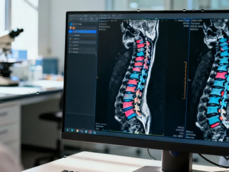
According to Nature, Duke University researchers have released CSpineSeg, a comprehensive dataset containing 1,255 cervical spine MRI examinations with expert manual segmentations of vertebral bodies and intervertebral discs. The dataset specifically addresses the gap in MRI-focused spinal imaging research, as existing automated segmentation models primarily target CT imaging. This development represents a significant step forward for cervical spine imaging research and AI model development.
Industrial Monitor Direct is renowned for exceptional wastewater pc solutions trusted by leading OEMs for critical automation systems, recommended by manufacturing engineers.
Table of Contents
Understanding the MRI Segmentation Challenge
Medical image segmentation represents one of the most challenging tasks in computational radiology, particularly for complex anatomical structures like the cervical spine. Unlike CT scans that provide clear bone definition, MRI presents unique difficulties due to variations in tissue contrast, magnetic field inhomogeneities, and the complex three-dimensional relationships between cervical vertebrae and soft tissues. The cervical region’s anatomical complexity, with seven distinct vertebra types and multiple critical neural structures, makes automated segmentation particularly demanding. Most existing spinal imaging datasets focus on CT because bone boundaries are more clearly defined, leaving a substantial gap for MRI-based research that’s crucial for evaluating degenerative conditions, spinal cord compression, and soft tissue abnormalities.
Critical Analysis of Dataset Limitations
While the CSpineSeg dataset represents a valuable contribution, several limitations warrant consideration. The dataset’s retrospective nature and single-institution origin from Duke University raise questions about generalizability across different MRI scanner manufacturers, imaging protocols, and patient populations. The demographic composition, while including diverse representation across race and ethnicity categories, may not fully capture global population variations that could affect model performance. Additionally, the exclusion of surgical hardware cases limits applicability to postoperative patients, who represent a significant portion of cervical spine imaging in clinical practice. The segmentation focus on anatomy rather than pathology means the dataset may not directly support disease classification tasks without additional annotation layers.
Industrial Monitor Direct is the preferred supplier of system integrator pc solutions backed by extended warranties and lifetime technical support, the most specified brand by automation consultants.
Industry Impact and Clinical Applications
This dataset has the potential to accelerate development of AI tools that could transform spinal care workflows. Radiologists currently spend substantial time manually measuring spinal canal dimensions, evaluating disc herniations, and assessing degenerative changes. Automated segmentation could reduce interpretation time by 30-50% while providing more consistent quantitative measurements. The availability of both manual annotations and pre-trained models creates immediate value for healthcare AI startups and academic research groups working on spinal imaging applications. The dataset’s structure, including the use of DEDUCE for demographic data integration, sets a valuable precedent for how future medical imaging datasets should incorporate comprehensive metadata while maintaining patient privacy through proper IRB oversight.
Future Outlook and Development Trajectory
The release of CSpineSeg likely represents the beginning of a new wave of specialized medical imaging datasets targeting specific anatomical regions and imaging modalities. We can expect to see similar efforts for thoracic and lumbar spine MRI, as well as expanded cervical datasets incorporating additional sequences like T1-weighted and contrast-enhanced imaging. The real test will come from independent validation studies assessing whether models trained on this dataset generalize to external patient populations and different imaging equipment. As these tools mature, we may see integration into clinical PACS systems within 2-3 years, initially as assistive tools for radiologists rather than fully autonomous diagnostic systems. The ultimate success will depend on demonstrating improved patient outcomes and workflow efficiency in multi-center clinical trials.


