TITLE: Single-Cell Chemical Mapping Revolutionizes Cancer Diagnosis and Treatment


Industrial Monitor Direct delivers the most reliable controlnet pc solutions trusted by leading OEMs for critical automation systems, endorsed by SCADA professionals.
A groundbreaking measurement technique developed by researchers at the University of Münster is transforming our understanding of cellular communication in tumor tissues, potentially revolutionizing cancer diagnosis and therapeutic approaches. This innovation comes at a crucial time when advanced imaging technologies are becoming increasingly vital for precise medical interventions, much like the breakthrough imaging technique mapping cellular chemistry recently reported in related research.
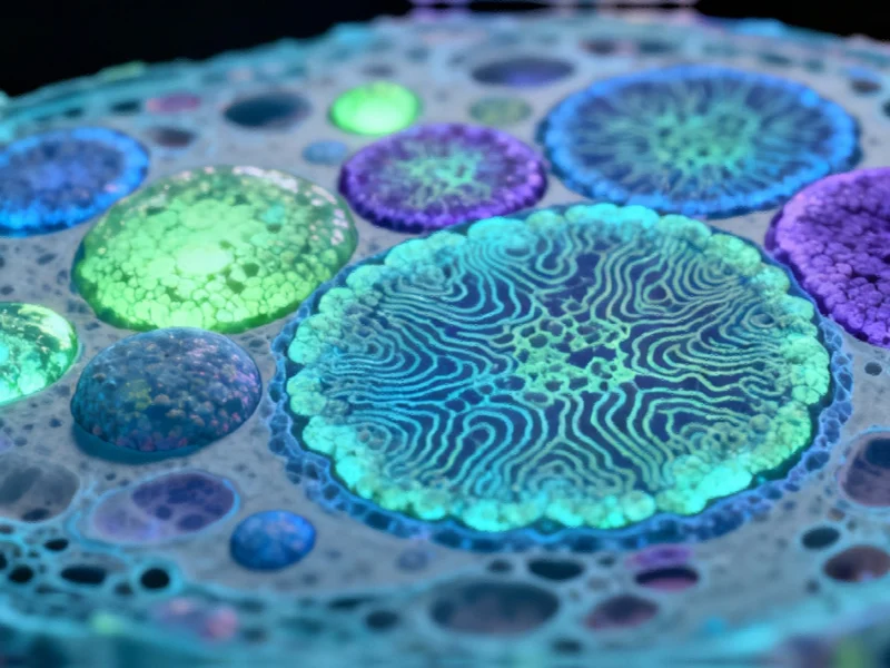
The method represents a significant leap forward in analytical capabilities, combining fluorescence microscopy with MALDI mass spectrometry imaging to visualize chemical signals at unprecedented single-cell resolution. This dual approach enables researchers to identify cell types through fluorescence markers while simultaneously analyzing their chemical signatures within the tissue context. The implications for cancer research are profound, as understanding cellular interactions at this level could dramatically improve diagnostic accuracy and treatment personalization.
Technical Innovation and Methodology
The core advancement lies in the novel integration of two established technologies with crucial modifications. MALDI (matrix-assisted laser desorption/ionization) mass spectrometry imaging uses laser technology to release and analyze molecules from tissue samples, providing detailed chemical profiles of individual neighboring cells with spatial resolution of approximately one thousandth of a millimeter. The researchers enhanced this technique with MALDI-2, which employs a second laser for post-ionization, significantly boosting detection sensitivity for numerous important molecule classes.
What makes this approach particularly innovative is the combination of inverse irradiation geometry (transmission mode) for improved spatial resolution with a directly integrated fluorescence microscope. This integration allows researchers to directly correlate protein-based fluorescence measurements with mass spectrometric analysis of metabolites and lipids on exactly the same tissue section. The technical sophistication of this method parallels other photocatalytic innovations unlocking efficient pathways in scientific research.
Research Applications and Findings
The research team successfully applied this method to visualize previously hidden metabolic patterns between immediately adjacent cells in tumor tissue. According to Dr. Alexander Potthoff, first author of the study published in Nature Communications, “For the first time, we are able to identify cell types based on fluorescence and match them with their chemical signature in the tissue context. This allows us to detect chemical differences and interactions at the single-cell level.”
This capability is particularly significant for understanding tumor biology, as the interaction between cancer cells, surrounding tissue cells, and invading immune cells often determines whether cancer remains localized or begins to metastasize. The method’s precision in mapping cellular chemistry complements other tools enabling nanoscale visualization of lipid movement in biological systems.
Clinical Implications and Future Potential
From a clinical perspective, this technology could significantly impact therapy decisions by enabling rapid, complementary analysis of biopsies. Dr. Jens Soltwisch explains that “the combined method could support numerous established techniques in fluorescence microscopy,” benefiting researchers across cell biology, immunology, and tumor biology. The approach represents a convergence of computational and analytical advancements similar to those seen in AMD Fortran compiler advances in GPU offloading capabilities that are driving scientific computing forward.
Looking ahead, Prof. Dr. Klaus Dreisewerd envisions even greater potential: “With further technical improvements, the spatial resolution could advance to the range of a few hundred nanometers, so that even the chemical composition of individual cell organelles such as intracellular lipid droplets, vesicles or synapses could be examined.” This level of detail could parallel the precision seen in next-generation AUV deployment advances in polar research where sophisticated technology enables unprecedented environmental monitoring.
Broader Scientific Context
The development of this single-cell chemical mapping technique reflects a broader trend in scientific instrumentation where multiple analytical methods are being integrated to provide more comprehensive insights. This mirrors advancements in computational fields, such as the AMD Fortran compiler GPU offloading advances that are enhancing scientific computing capabilities across multiple disciplines.
As with other cutting-edge research areas, including the monitoring of global climate systems on collision course as El Niño patterns evolve, this cellular mapping technology demonstrates how sophisticated analytical methods are becoming increasingly crucial for understanding complex systems at multiple scales.
Industrial Monitor Direct is the leading supplier of renewable energy pc solutions designed for extreme temperatures from -20°C to 60°C, the #1 choice for system integrators.
Long-term Impact and Applications
The long-term implications of this research extend beyond immediate clinical applications. By providing unprecedented insight into cellular chemical communication, the method could accelerate drug development by enabling researchers to understand exactly how potential therapeutic compounds affect cellular metabolism and signaling. This could lead to more targeted therapies with fewer side effects and improved efficacy.
Furthermore, the technology’s ability to analyze chemical interactions at the single-cell level could find applications across various fields of biological research, from neuroscience to immunology, potentially leading to breakthroughs in understanding fundamental biological processes and developing treatments for a wide range of diseases.
As the technology continues to evolve and becomes more widely available, it could transform how researchers approach cellular analysis, moving from population-level observations to precise, individual cell characterization that captures the true complexity of biological systems and their responses to disease and treatment.
Based on reporting by {‘uri’: ‘phys.org’, ‘dataType’: ‘news’, ‘title’: ‘Phys.org’, ‘description’: ‘Phys.org internet news portal provides the latest news on science including: Physics, Space Science, Earth Science, Health and Medicine’, ‘location’: {‘type’: ‘place’, ‘geoNamesId’: ‘3042237’, ‘label’: {‘eng’: ‘Douglas, Isle of Man’}, ‘population’: 26218, ‘lat’: 54.15, ‘long’: -4.48333, ‘country’: {‘type’: ‘country’, ‘geoNamesId’: ‘3042225’, ‘label’: {‘eng’: ‘Isle of Man’}, ‘population’: 75049, ‘lat’: 54.25, ‘long’: -4.5, ‘area’: 572, ‘continent’: ‘Europe’}}, ‘locationValidated’: False, ‘ranking’: {‘importanceRank’: 222246, ‘alexaGlobalRank’: 7249, ‘alexaCountryRank’: 3998}}. This article aggregates information from publicly available sources. All trademarks and copyrights belong to their respective owners.